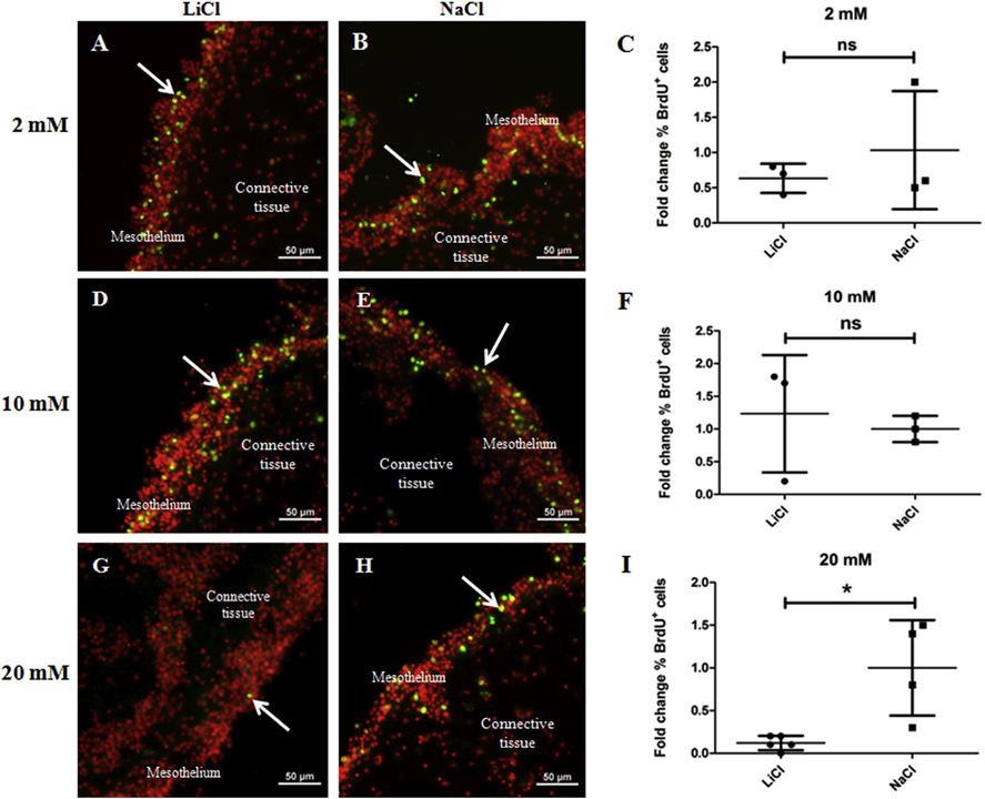
Figure 10. 20 mM LiCl reduces mesothelial cell proliferation in gut rudiment explants from 14 dpe animals.
Cell proliferation was assayed by BrdU incorporation. Representative histological cross sections of explants showing the effect of LiCl at 2 mM (A), 10 mM (D), and 20 mM (G) and their respective controls: 2 mM (B), 10 mM (E), and 20 mM (H) NaCl on mesothelial cell proliferation. LiCl doses of 2 mM and 10 mM did not have significant effects on the percentage of BrdU labeled cells in the mesothelium compared to controls (2 mM and 10 mM NaCl-treated explants) (C and F, respectively). However, 20 mM LiCl treatment resulted in a significant reduction in the percentage of BrdU+ cells in the mesothelium compared to controls (20 mM NaCl-treated explants) (I). Results represent the mean ± SD, t-test, * p<0.05, n = 3 in 2mM and 10 mM LiCl, n = 3 in 2 mM and 10 mM NaCl, n = 5 in 20 mM LiCl, n = 4 in 20 mM NaCl. Cell nuclei are shown in red (DAPI) and BrdU labeled nuclei (white arrows) in green. Scale bar = 50 μm.

