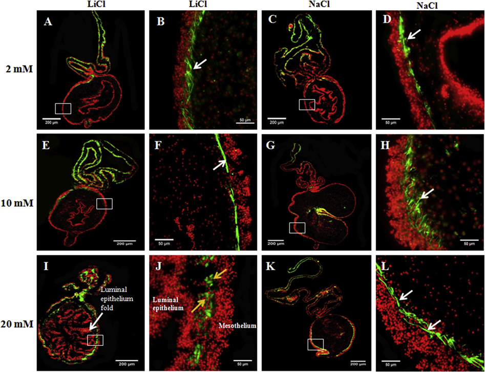
Figure 11. 20 mM LiCl induces muscle dedifferentiation in gut rudiment explants from 14 dpe animals.
The ability of LiCl at three doses (2, 10, and 20 mM) to promote gut rudiment muscle dedifferentiation was tested in explants from 14 dpe. Representative histological cross sections of intestines and attached mesenteries showing the effect of LiCl at 2 mM (A,B), 10 mM (E,F), and 20 mM (I,J) and their respective controls: 2 mM (C,D), 10 mM (G,H), and 20 mM (K,L) NaCl. Explants treated with 2 mM LiCl (B) or 10 mM LiCl (F) showed a muscle layer similar to those observed in control explants, 2 mM NaCl (D) and 10 mM NaCl (H). Moreover, these explants had similar morphology (A and E, respectively) to control explants (C and G, respectively). However, explants treated with 20 mM showed SLSs and small spherical cells instead of a defined muscle layer (J). These explants also presented altered tissue morphology with abundant luminal epithelium folds and a reduced connective tissue space (I). Conversely, control explants (treated with 20mM NaCl) showed a defined muscle layer in the gut rudiment (L) and typical explant morphology (K). The micrographs on the first (A,E,I) and third column (C,G,K) show the LiCl and NaCl-treated explant morphology, respectively, at low magnification. The micrographs on the second and fourth columns show the gut rudiment muscle layer (white arrows) or SLSs (yellow arrows) at higher magnification in LiCl and NaCl-treated explants, respectively, in green. Cell nuclei (DAPI) are shown in red. The mesothelium and luminal epithelium are pointed out in 20 mM LiCl treated explants (I,J). n = 4. Scale bar = 200 μm (A,E,I,C,G,K) and 50 μm (B,F,J,D,H,L).

