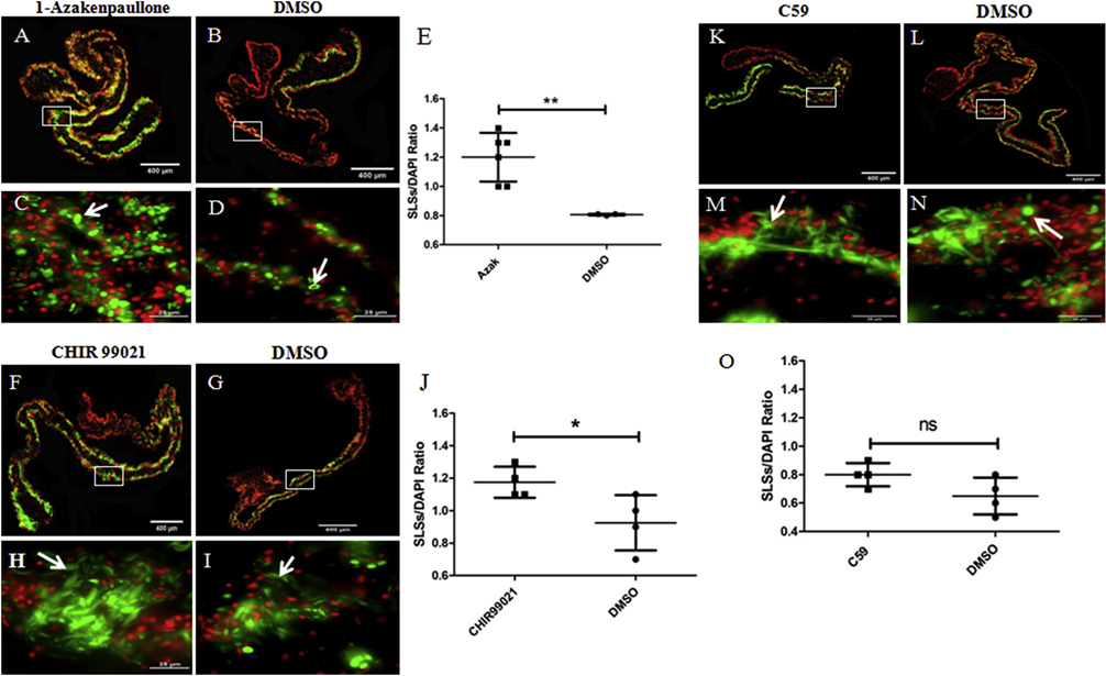
Figure 12. Effects of known GSK-3 inhibitors and C59 on muscle dedifferentiation in vitro.
The SLSs/DAPI ratios in explants treated with the GSK-3 inhibitors 1-Azakenpaullone and CHIR99021, and the Wnt antagonist C59, were measured as indicators of muscle dedifferentiation. Representative histological cross sections of explants showing the effect of 1-Azakenpaullone (A,C), CHIR99021 (F,H), C59 (K,M) and their controls (B,D), (G,I), and (L,N), respectively. The micrographs on the bottom (C,D,H,I,M,N) are high-magnification view of the boxed regions in the top micrographs (A,B,F,G,K,L respectively). Both 1-Azakenpaullone (E) and CHIR99021 treatment (J) increased significantly the SLSs/DAPI ratio compared to controls. Conversely, we did not find significant differences in the SLS/DAPI ratio between C59 treated explants and their controls (DMSO) (O). Results represent the mean ± SD, t-test, **p<0.01, *p<0.05, n = 6 Azakenpaullone, n = 4 CHIR99021, n = 4 C59, n = 3–4 DMSO. SLSs (white arrows) are shown in green (phalloidin-TRITC) and cell nuclei in red (DAPI). Scale bar = 400 μm (A,B,F,G,K,L), 25 μm (C,D,H,I,M,N).

