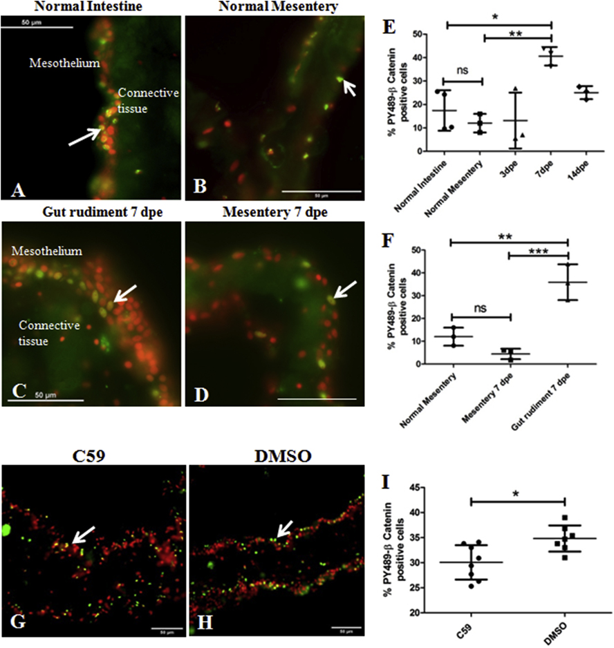
Figure 14. In vivo and in vitro expression of PY489-β-catenin.
Activation of the Canonical β-catenin pathway in normal and regenerating intestines was determined in vivo (A-F) and in vitro (G-I) by immunohistochemistry using the monoclonal antibody Anti-PY489-β-catenin to detect its nuclear expression, a proxy for β -catenin activation. Representative histological cross sections of normal intestinal mesothelium (A), normal mesentery (B), gut rudiments at 7 dpe (C) and regenerating mesentery at 7 dpe (D) show the nuclear labeling of the antibody to be mainly in some mesothelial cells. A significant increase in the percentage of mesothelial cell nuclei labeled with the PY489-β-catenin antibody was observed at 7 dpe compared to the normal intestine and mesentery (E). The increase in PY489-β-catenin immunoreactive cell nuclei was localized to the gut rudiment; no significant differences in the percentage was observed between normal mesentery and regenerating mesentery at 7 dpe (F). Results represent the mean ± SD, One-Way ANOVA, * p<0.05, *p<0.01, n = 3–4. Treatment of in vitro explants with C59 reduced nuclear β-catenin expression. Representative histological cross sections of gut explants treated with C59 (G) and its control (DMSO) (H). C59 caused a significant reduction in the percentage of PY489-β-catenin labeled cell nuclei compared to controls (DMSO), suggesting that this drug inhibits the Wnt pathway in gut explants (I). Results represent the mean ± SD, t-test, * p<0.05, n = 8 for C59, n = 7 for DMSO. Cell nuclei are shown in red (DAPI) and PY489-β-catenin labeled nuclei (white arrows) in green. Scale bar = 50 μm.

