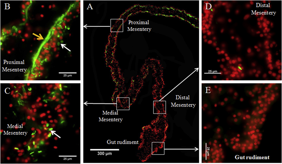
Figure 3. Spindle-like structures (SLSs) and muscle fibers gradient in regenerating intestines in H. glaberrima.
Representative histological cross sections of a gut rudiment and the attached mesentery showing the gradient of SLSs and muscle fibers observed during early stages of intestine regeneration. The whole gut rudiment and adjacent mesentery is shown in (A). At higher magnification are shown the proximal (B), medial (C), and distal mesentery (D), as well as the gut rudiment (E). Muscle fibers (yellow arrows) are observed in proximal mesentery and SLSs (white arrows) are observed in both proximal and medial mesentery. Conversely, no SLSs or muscle fibers are present in distal mesentery and gut rudiment. SLSs and muscle fibers are shown in green (Phalloidin-TRITC) and cell nuclei in red (DAPI). Scale bars = 300 μm in (A) and 25 μm in (B), (C), (D), and (E).

