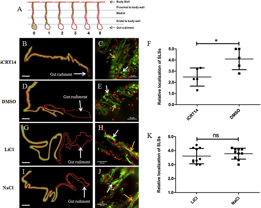
Figure 4. Effect of iCRT14 on mesentery muscle dedifferentiation in vivo.
(A) Diagram that depicts the staging system used for quantifying mesenterial muscle dedifferentiation in vivo. Stages are based on the localization of the SLSs in the mesentery relative to the body wall and the gut rudiment. Representative histological cross sections of gut rudiments and attached mesenteries showing the effects of iCRT14 (B) and LiCl (G) treatments on mesenterial muscle dedifferentiation compared to controls DMSO (D) and NaCl (I), respectively are shown. The areas of the mesentery where SLSs were observed in the different tissues are presented in green in the schemes for clarity purposes (B,D,G,I). SLSs were observed adjacent to the gut rudiment in iCRT14-treated animals (B) while they were localized in the middle segment of the mesentery in control animals (DMSO-treated) (D). Furthermore, SLSs were observed in the middle segment of the mesentery in both LiCl (G) and NaCl (I) treated groups. The micrographs on the right (C,E,H,J) show the presence of SLS (white arrows) and muscle fibers (yellow arrows) in the different samples. A significant difference in the localization of SLSs was found between iCRT14 -treated animals and controls, suggesting a delay in muscle dedifferentiation due to iCRT14 treatment (F). Conversely, no significant differences were found between LiCl and control (K). Results represent the mean ± SD, t-test, *p<0.05, n= 5 iCRT14, n =5 DMSO, n= 10 LiCl, n= 10 NaCl. SLSs and muscle fibers are shown in green (Phalloidin-TRITC) and cell nuclei in red (DAPI). The gut rudiment is pointed out in the schemes of the tissue sections. The schemes were drawn to scale. Scale = 300 μm (B,D,G,I) and 25 μm (C,E,H,J).

