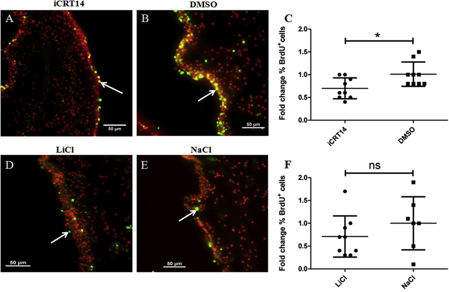
Fig 5. iCRT14 but not LiCl alter cell proliferation during intestine regeneration in vivo.
Cell proliferation was assayed by BrdU incorporation. Representative histological cross sections of gut rudiments showing the effect of iCRT14 (A) and LiCl (D) treatments on cell proliferation compared to controls DMSO (B) and NaCl (E), respectively. iCRT14 reduces significantly the percentage of BrdU labeled cells compared to controls (DMSO) (C). However, no significant differences in the percentage of BrdU labeled cells were found between LiCl-treated animals and their controls NaCl (F). Results represent the mean ± SD, t-test, *p<0.05, n = 9 iCRT14, n = 9 DMSO, n = 9 LiCl, n = 7 NaCl. Cell nuclei are shown in red (DAPI) and BrdU+ nuclei (white arrows) in green. Scale bar = 50 μm.

