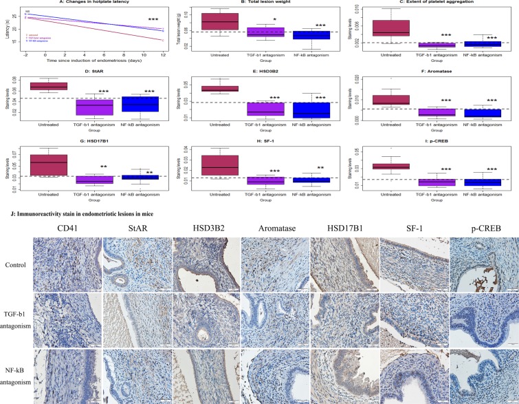
Figure 7.
Effect of antagonism of TGF-β1 (by SB431542) or of NF-κB (by JSH-23) on lesional development and lesional expression of steroidogenic proteins in mice with induced endometriosis. (A) Change in hotplate latency for mice treatment with vehicle (untreated, or U), antagonism of TGF-β1 (T), and antagonism of NF-κB (N) tested at the time indicated. (B) Total lesion weight in the 3 groups of mice. (C) Boxplots of the extent of lesional platelet aggregation using CD41 the 3 groups of mice. Boxplots of lesional staining levels in the 3 groups of mice: StAR (D), HSD3B2 (E), aromatase (F), HSD17B1 (G), SF-1 (H), and p-CREB (I). The dotted horizontal line represents the median of all 3 groups. Symbols of statistical significance: *: p < 0.05, **: p < 0.01, ***: p < 0.001. NS: p > 0.05. n = 10 for each group. Except in (A), where the comparison was made using Kruskal’s test, all tests were made using Wilcoxon’s test in comparison with the untreated group (U). (J) Representative immunoreactivity staining of CD41, StAR, HSD3B2, aromatase, HSD17B1, SF-1 and p-CREB in the endometriotic lesions (×400) in Balb/c mice with induced endometriosis which then treatment with vehicle, SB431542 (an antagonist for TGF-β1) and JSH-23 (an antagonist for NF-κB) for two weeks. Scale bar = 50 μm.

