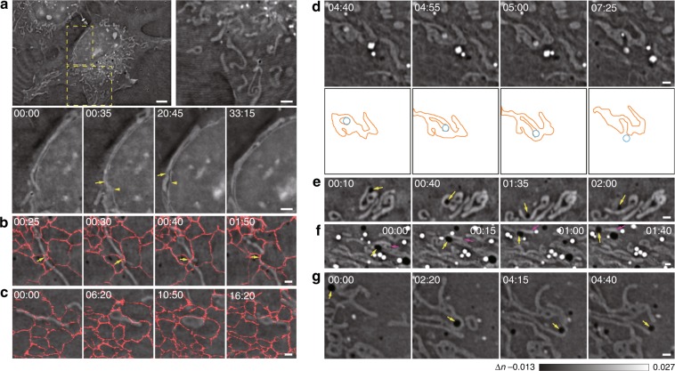
Fig. 4. Different dynamics of mitochondria interacting with other organelles.
a Representative examples of different shapes of cytosolic mitochondria and mitochondria associated with the nuclear membrane. In contrast to cytosolic mitochondria that have various shapes, lengths, and orientations (enlarged in the right panel), long mitochondria associated with the nuclear membrane for half an hour (enlarged and shown as a montage in the lower panel) and exhibited dynamic morphological changes with reciprocal movement of the nuclear membrane (arrows and arrowheads). b, c Two representative distinct examples of ER–mitochondria contacts. One contact (indicated by the arrows) led to scission of the mitochondrion (b), while the other contact led to the lateral expansion of the mitochondrion (c). We overlaid the fluorescent signals of KDEL-EGFP on these images to better present the data. d A representative example of a lysosome that was initially surrounded by the same mitochondrion, which caused fission and then moved out of the mitochondrion. Montages of actual images (top) and corresponding schematic diagrams (bottom) are shown. e–g Representative examples of three modes of mitochondrion–DB contacts. The collision of DBs (arrows) with the mitochondrion changed the mitochondrial morphology (e), caused the mitochondrion (magenta arrows) to move along with the DB (highlighted by yellow arrows in f), or caused fission (arrows) of the interacting mitochondrion (g). Scale bars: (a) 5 μm and 2 μm (zoom in); (b–g) 2 μm.

