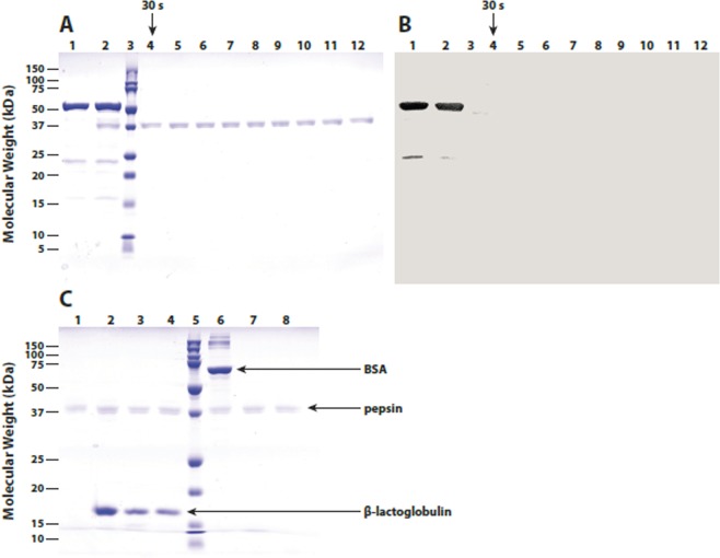
Figure 4.
Panels A and B: Samples of CRTI protein purified from recombinant E. coli (Lot No. M20454-02) were incubated in the presence of SGF pH 1.2 containing pepsin for 0 min (lane 2) and 0.5, 1, 2, 5, 10, 20, 30 or 60 min at 37 °C (lanes 4–11) and then analyzed by SDS-PAGE. Gels were either stained for protein with colloidal blue G250 (panel A) or subjected to western immunoblot analysis (panel B) using rabbit anti-CRTI immunoglobulin (1:1000) and horseradish peroxidase-conjugated goat anti-rabbit IgG followed by precipitating substrate development. Control samples included CRTI protein diluted in gastric control fluid without pepsin (lane 1) and SGF solution containing pepsin (lane 12). Molecular weight standards are shown in lane 3 (adapted from GR2E-FFP submitted study reports).

