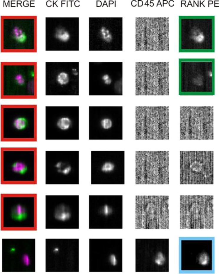
Figure 7.
RANK immunostaining of CTCs ex vivo. Analysis of five rare cells and one leukocyte in a blood sample of a breast cancer patient using an Analyzer II device. Horizontally, the photos show the same cell stained for the combination (DAPI/CK FITC) of CK (green) and DAPI (violet); CK FITC only; DAPI only; CD45 APC only; and RANK PE only. The red squares indicate positively stained cells: based on RANK staining profile (sufficient signal relative to background) we classified the first two cells as RANK-positive CTCs (green squares). Based on CD45 expression, the bottom photos show a leukocyte that express RANK (light blue square), and serve as internal fluorescence control.

