Figure 2.

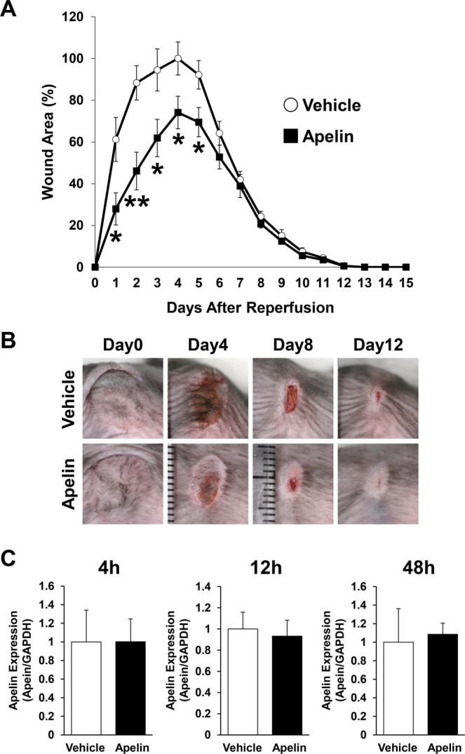
Apelin protected PUs formation in cutaneous I/R injury mouse model. (A) The size of the wound area after I/R injury in normal C57BL/6 mice treated with subcutaneous injection of apelin (10 ng/mice) or phosphate-buffered saline as a control. The size of the ulcer in control mice at 4 days after reperfusion was assigned a value of 100% (vehicle: n = 9, apelin: n = 10, for each time point and group). (B) Representative images of the wound after cutaneous I/R in control or apelin treated mice at 0, 4, 8, and 12 days after reperfusion. (C) Comparison of mRNA levels of apelin expression during wound healing in I/R site between control and apelin-treated mice at 4, 12 and 48 hours after I/R injury. n = 5. All values represent mean ± SEM. **P < 0.01, *P < 0.05.
