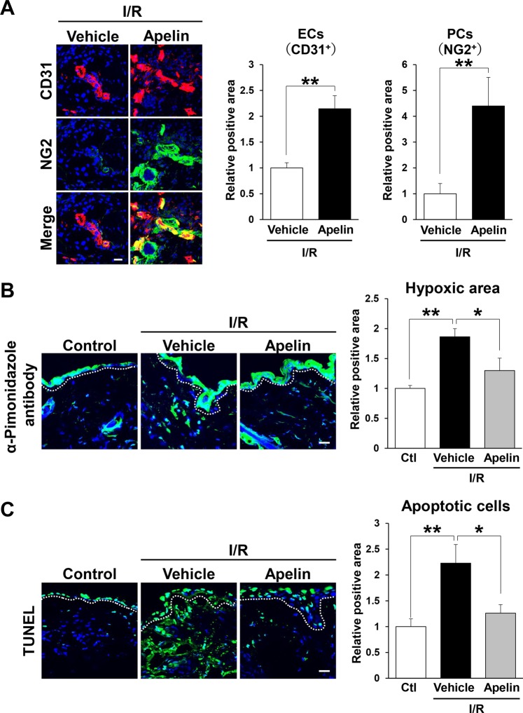
Figure 3.
Apelin protected vascular reduction and suppressed hypoxia and apoptosis after cutaneous I/R. (A) The amount of CD31+ ECs and NG2+ pericytes in the cutaneous I/R area at 4 days after reperfusion. (B) The amount of pimonidazole+ hypoxic area in cutaneous I/R site at 1 day after reperfusion. Quantification of the pimonidazole+ areas in 6 random microscopic fields from the center of I/R area in n = 3 mice per groups was performed using ImageJ software. Positive area in control mice was assigned a value of 1. Scale bar = 20 μm. (C) The number of apoptotic cells in the I/R site at 1 day after reperfusion was determined by counting both TUNEL- and DAPI-positive cells. Values were determined in 6 random microscopic fields in n = 3 mice per group. All values represent mean ± SEM. **P < 0.01, *P < 0.05.

