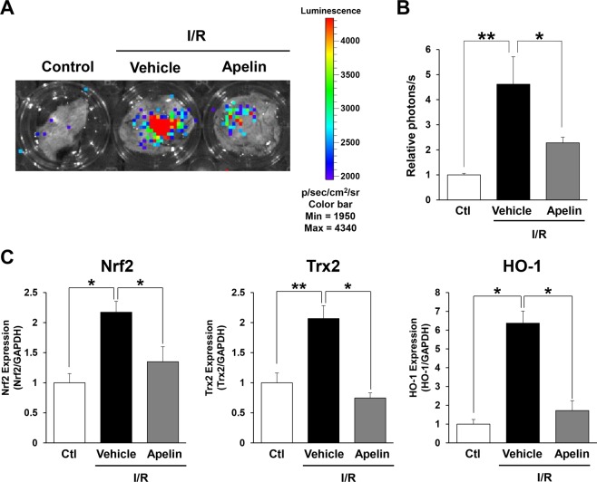
Figure 5.
Apelin reduced oxidative stress induced by cutaneous I/R injury in vivo. (A) Representative image of luminescence signals in cutaneous I/R area in OKD48 mice at 24 hours after reperfusion (Day 1). The color scale bar shows the photon counts (photon (p)/sec/cm2/sr). (B) Quantification of luminescence signals in cutaneous I/R area in OKD 48 mice. n = 4 in each group. (C) mRNA levels of oxidative stress-associated factors, Nrf2, Trx2 and HO-1 in the I/R area at 24 hours after reperfusion (Day 1). mRNA levels in control mice were assigned as values of 1. Values represent mean ± SEM. n = 4–5 mice in each group. **P < 0.01, *P < 0.05.

