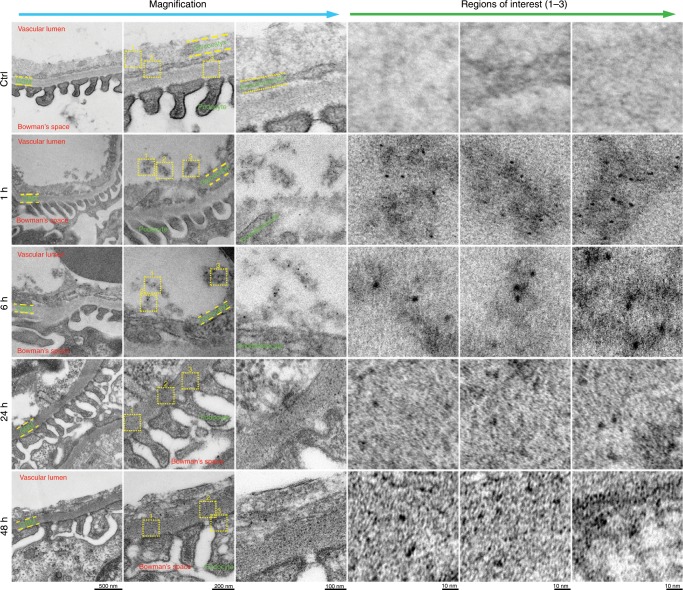
Fig. 3. Glomerulus ultrastructure after cisplatin treatment.
No NPs were observed in the region of the glomerular filtration membrane, including the glycocalyx, endothelial cells and GBM (n = 10), in the control mice treated with PBS (n = 10). In the cisplatin-treated mice, a large number of small Pt NPs (<6 nm) were observed at 1 h, while larger Pt NPs (>6 nm) were observed at 6 h. The Pt NPs (>6 nm) were found in the GBM region at 24 h and 48 h, indicating a slow elimination through the glomerulus. Regions of interest are shown at a higher magnification.

