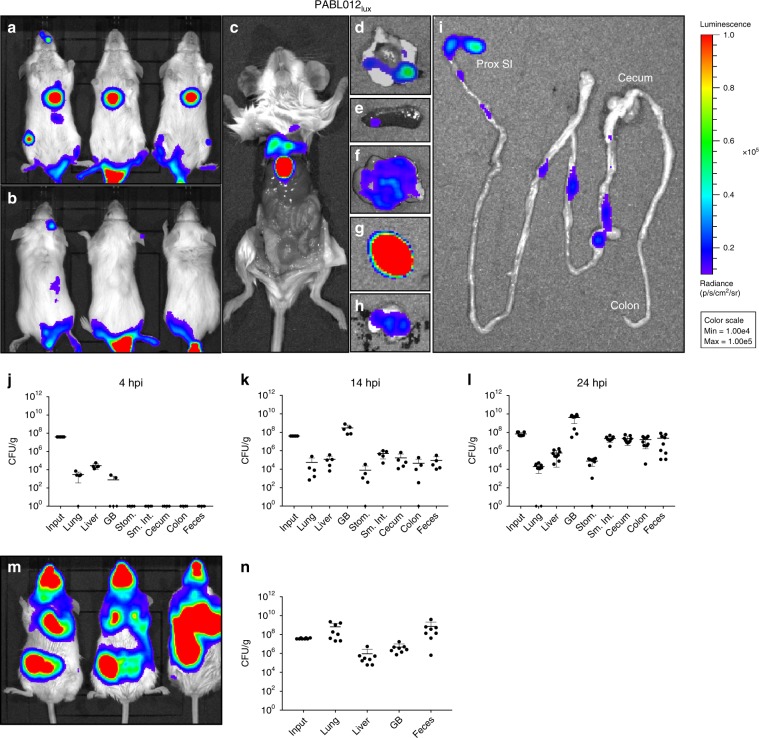
Fig. 1. P. aeruginosa disseminates to the gallbladder and intestines following bacteremia and pneumonia.
Mice (n = 3, representative replicate) were intravenously injected with ~2 × 10⁶ CFU of a luciferase expressing strain of P. aeruginosa, PABL012lux. Ventral (a) and dorsal (b) views of live mice at 24 hour post infection (hpi) were obtained using IVIS. Mice were subsequently euthanized, the abdominal wall dissected away and internal organs visualized (c). The heart and lung (d), spleen (e), liver (f), gallbladder (g), stomach (h), and remaining intestinal tract (i) of a single mouse were separately removed and imaged to visualize bacterial localization. (Since bioluminescence is dependent on oxygen, the signal present in the anaerobic portions of the intestinal tract may underestimate bacterial numbers.) The scale (red = high, blue = low) for all images represents radiance (photons per sec per cm2 per steradian) with minimum and maximum values normalized to 1 × 10⁴ and 1 × 10⁵, respectively. Intravenously infected mice housed on standard bedding were euthanized at 4 hpi (j, n = 5), 14 hpi (k, n = 5), or 24 hpi (l, n = 3 × 3 replicates). Bacteria were enumerated from several anatomical sites by plating serial dilutions of organ homogenates. m BALB/c mice (n = 3, representative replicate) were infected with PABL012lux by intranasal inoculation of ~2 × 10⁶ CFU and imaged at 24 hpi using IVIS. n Mice (n = 4 × 2 replicates) were subsequently euthanized and bacteria were enumerated from several anatomical sites. Density (CFU per gram [CFU/g] of organ weight) of recovered bacteria are presented (black circles). Organs where no bacteria were recovered are denoted with a diamond on the x-axis. Geometric means (horizontal lines) and SD (whiskers) are shown. ProxSI (proximal small intestine), GB (gallbladder), Stom. (stomach), Sm.Int. (small intestine).

