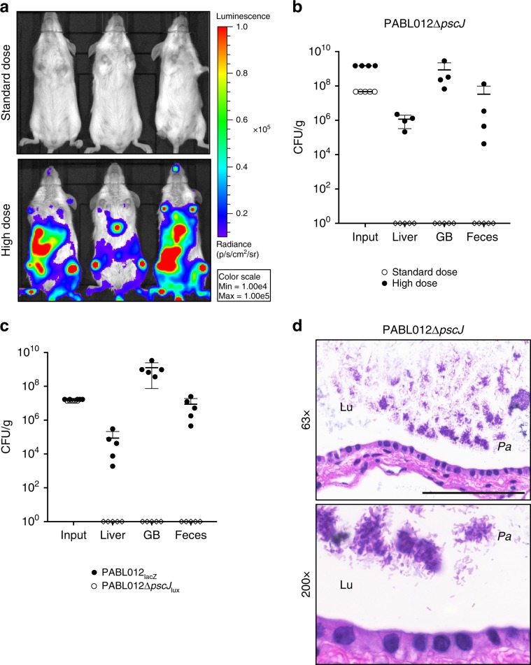
Fig. 5. The P. aeruginosa type III secretion system (T3SS) contributes to gastrointestinal shedding.
a BALB/c mice (n = 3, representative replicate) were infected intravenously with a standard (~2 × 10⁶ CFU, open circles) or a high dose (~8 × 10⁷ CFU, filled circles) of a PABL012∆pscJlux T3SS mutant and imaged at 24 hpi using IVIS. The scale (red = high, blue = low) for all images represents radiance (photons per sec per cm2 per steradian) with minimum and maximum values normalized to 1 × 10⁴ and 1 × 10⁵, respectively. b Mice (standard dose n = 5, open circles; high dose n = 4, black circles) were euthanized, bacteria enumerated from the liver, gallbladder (GB), and feces, and CFU per gram organ reported at 24 hpi. c BALB/c mice (n = 5) were co-infected intravenously with a 1:1 mix totaling ~2 × 10⁶ CFU of PABL012lacZ (n = 5, filled black circles) and PABL012∆pscJlux (n = 5, open symbols). Liver, GB, and feces were harvested at 24 hpi for enumeration of viable bacteria. For input 1 g equals 1 mL of wet weight. For scatter plots, each circle or diamond represents data from one mouse. Where no CFU were recovered, data are represented as diamonds on the x-axis. Geometric means (horizontal lines) and SD (whiskers) are shown. d H&E stained sections of gallbladders from mice infected with the high dose of PABL012∆pscJlux. Lu (lumen), Pa (P. aeruginosa). Scale bar represents 100 µM (×63).

