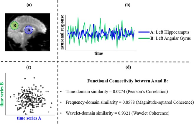
Figure 1.
A contrary case. Note: An example of FC within the standard DMN in a young healthy adult: (a) left hippocampus (denoted by A) and left angular gyrus (denoted by B) within the DMN are considered based on the Willard functional atlas; (b) the BOLD time series signals from preprocessed resting-state functional MRI were extracted from each region; (c) a scatter plot comparing the two BOLD time series shows the temporal linear correlation between them; (d) three distinct similarity measures of FC between the signals are compared; FC = functional connectivity; DMN = default mode network; BOLD = blood-oxygen-level-dependent;.

