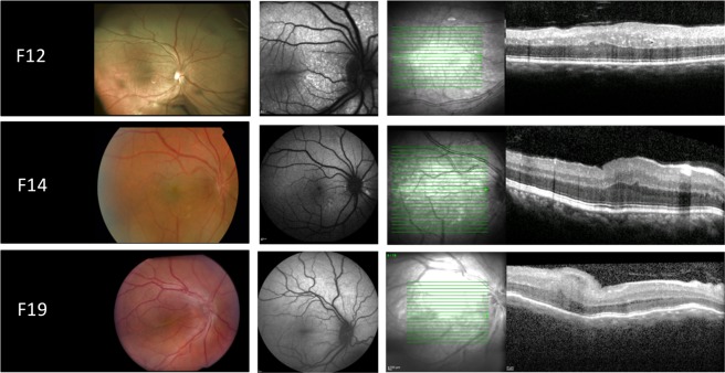
Figure 3.
Retinal imaging from the right eye of patients with NNO or MCOP due to mutations in PRSS56. Left panel: color fundus. Middle panel: fundus autofluorescence imaging (FAF). Right panel: optical coherence tomography (OCT). F12: papillomacular fold with diffuse white dots in the posterior pole and to a lesser extent throughout retina, increased autofluorescence at sites of white dots, thickened OCT with occasional intraretinal cyst. F14: papillomacular fold, large posterior pole white dots that have increased autofluorescence, thickened OCT. F19: color fundoscopy showing crowded optic disc and prominent papillomacular fold, normal autofluorescence on FAF imaging, thickened OCT with foveal hypoplasia and papillomacular fold.

