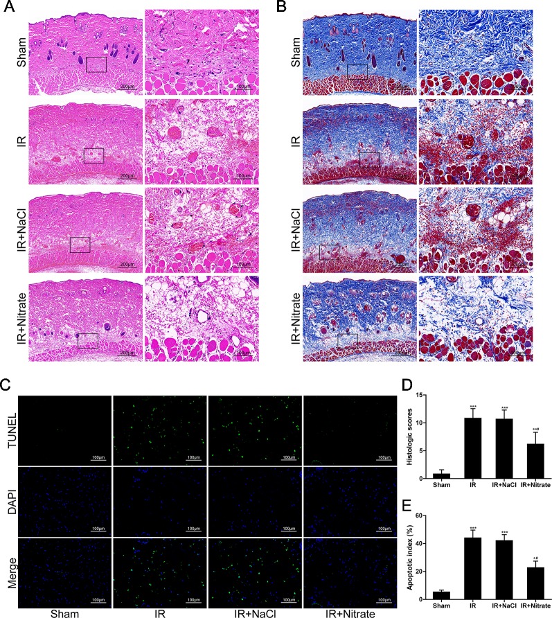
Figure 2.
Dietary nitrate decreased histological lesions and protected cells from apoptosis. (A, B) H&E and Masson trichrome staining of the musculocutaneous flap. (Large image, scale bar = 200 µm; small image, scale bar = 100 µm). (C) Representative TUNEL-stained images of the skin flap. The apoptotic cells were detected by TUNEL (green), and the nuclei were detected by DAPI (blue). Scale bar = 100 µm. (D) Quantitative analysis of the histologic scores. (E) Apoptotic index of the flap. Data are presented as mean ± SD, n = 6. *p < 0.05, **p < 0.01, ***p < 0.001 versus Sham group; #p < 0.05 versus IR group.

