
Fig. 2.
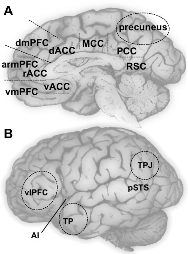
Brain regions of interest for self-development. Panel A displays a medial view of the brain with approximate locations of dorsal, anterior rostral, and ventral medial prefrontal cortex (dmPFC, armPFC, and vmPFC, respectively); dorsal, rostral, and ventral anterior cingulate cortex (dACC, rACC, and vACC, respectively); middle cingulate cortex (MCC); and medial posterior parietal cortex (mPPC) which includes precuneus, posterior cingulate cortex (PCC) and retrosplenial cortex (RSC). Panel B displays a lateral view of the brain with approximate locations of tempo–parietal junction (TPJ), posterior superior temporal sulcus (pSTS), temporal poles (TP), anterior insula (AI, which is underneath the lateral surface), and ventral lateral prefrontal cortex (vlPFC). Ventral striatum (VS), a subcortical structure, is not pictured.