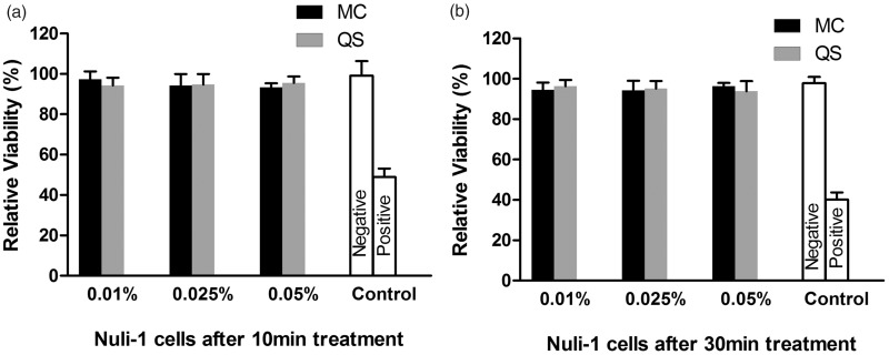
Figure 5.
Cell viability monitored by the LDH assay after 10-min (a) and 30-min (b) treatment of cultured human airway epithelial (Nuli-1) cells with CPC-quatsome and CPC micelle solutions. 2% Triton x-100 and 5% glucose in bronchial epithelial cell growth medium was used as the positive and negative control, respectively. The results were from two independent measurements with n = 6. The histogram displayed medians with ranges, and the comparison was carried out by Mann–Whitney test. MC: CPC micelle solution; QS: CPC-quatsome.

