
Figure 1.
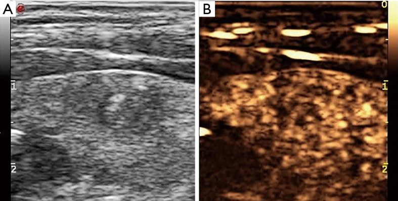
US of a thyroid nodule in a 38-year-old male who underwent fine-needle aspiration. Longitudinal US images demonstrating a hypoechoic nodule in the left thyroid gland (A). The nodule was hypoenhanced on CEUS (B). The cytology was papillary carcinoma and BRAF V600E (+). CEUS, contrast-enhanced ultrasound.