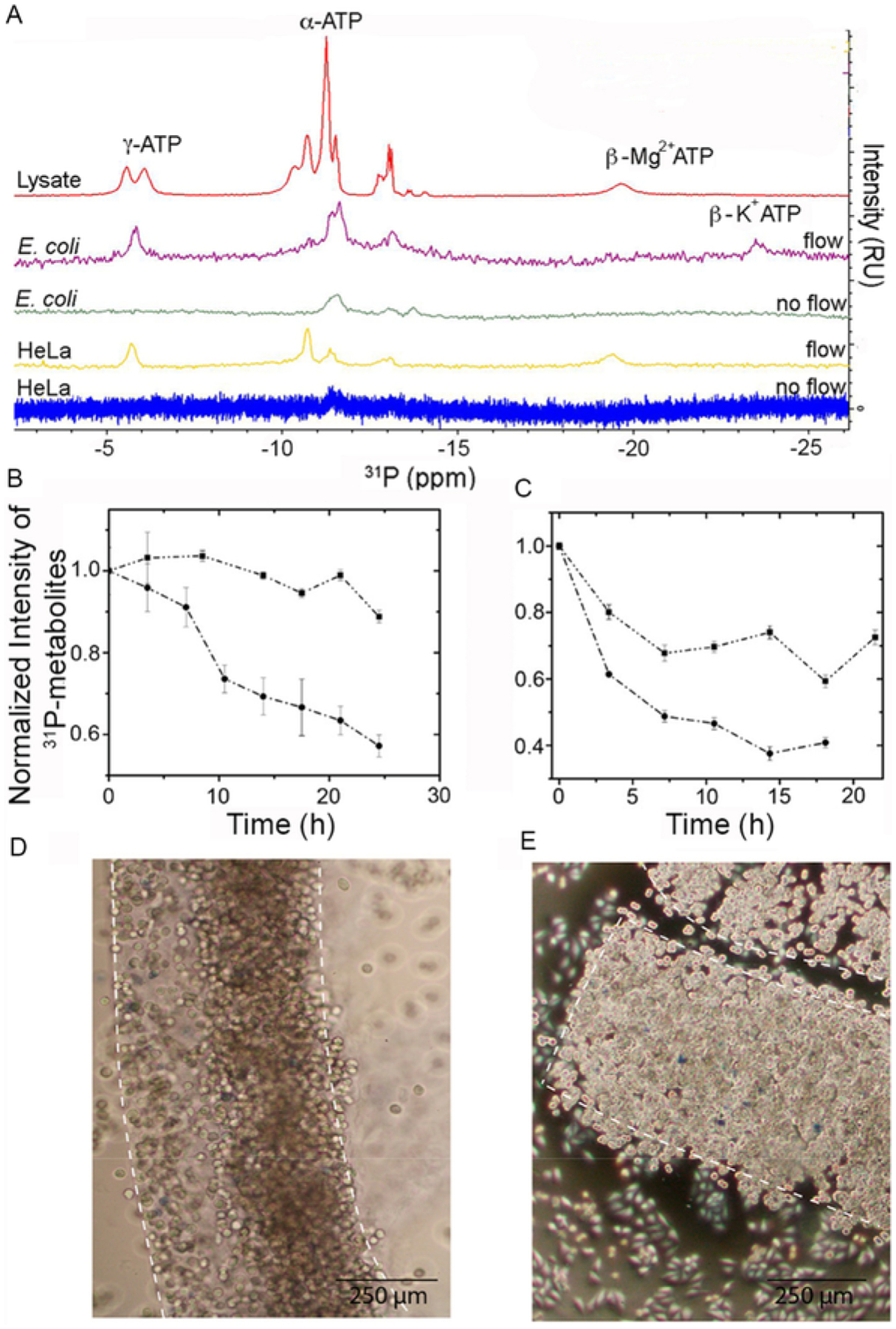
Fig. 4.

Cell viability is maintained for up to 24 h in a bioreactor. (A) 31P NMR spectra showing α-, β- and γ-ATP levels after 6 h for E. coli and HeLa cells in the bioreactor and in E. coli cell lysate. (B) Signal intensity of 31P-metabolites over time in E. coli with (squares) and without (circles) M9 flow. (C) Intensity of 31P-metabolites over time in HeLa cells with (squares) and without (circles) DMEM flow. (D) Trypan blue staining of cast HeLa cells before in-cell spectroscopy. (E) Trypan blue staining of cast HeLa cells 24 h after casting.
