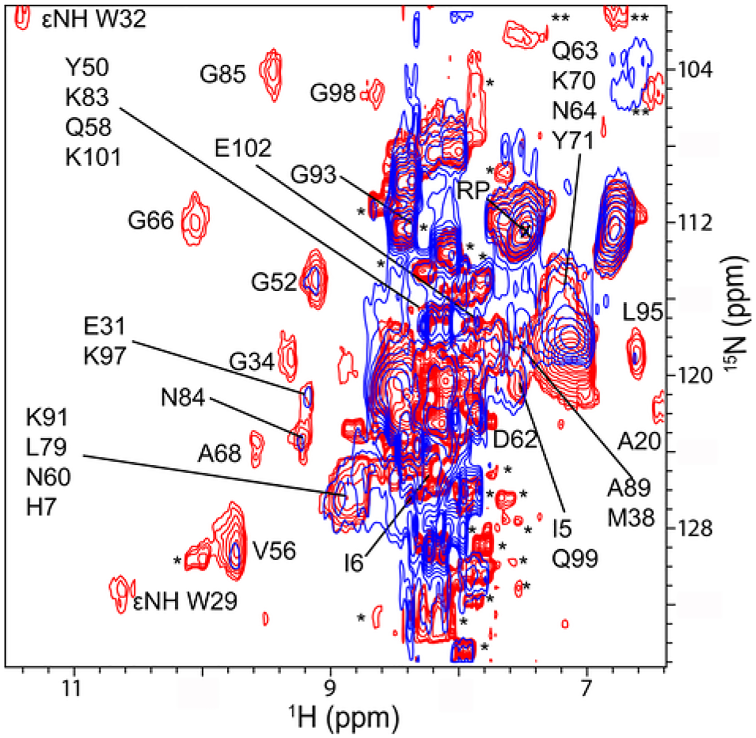
Fig. 5.

Binding of streptomycin to ribosomes changes the quinary structure of Trx in E. coli. Overlay of in-cell 1H −15N CRINEPT − HMQC − TROSY spectra of Trx without (red) and with (blue) streptomycin (final spectrum). Residues that broaden in the presence of streptomycin are indicated. Single and double asterisks indicate peaks from metabolites and unassigned side chain protons, respectively. The spectra are shown at the same contour levels. The reference peak used for intensity normalization is indicated by RP.
