
Figure 4.
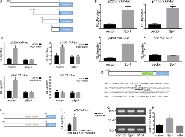
Sp‐1 (specificity protein 1) increased YAP (Yes‐associated protein) promoter activity in the process of SMC phenotypic modulation. A, Schematic showing the series of YAP promoter‐reporter plasmids used. B, Smooth muscle cells (SMCs) were transfected with the 2200‐, 1150‐, 450‐, or 50‐bp YAP promoter‐luciferase (Luc) plasmid and further transfected with vector or Sp‐1 plasmid before determining relative changes in luciferase expression. Data indicated that Sp‐1 overexpression‐mediated increases in YAP promoter activity required sequences between −450 and −50 bp. *P<0.05 vs vector group. n=3. C, siRNA knockdown of Sp‐1 abolished platelet‐derived growth factor‐BB (PDGF‐BB)‐induced activation of the YAP promoter. SMCs were cotransfected with the 2200‐, 1150‐, 450‐, or 50‐bp YAP promoter reporter plasmid and either control siRNA plasmid or siSp‐1 for 36 h, and treated with PDGF‐BB (20 ng/mL) or vehicle for 4 h before demonstrating relative changes in luciferase expression. *P<0.05 vs vehicle group. n=3. D, Sequence of the YAP promoter region from −450 to −50 showing the 2 consensus Sp‐1 binding sites. E and F, Mutation of the 2 Sp‐1 binding sites with the 450‐bp YAP promoter abolished basal and PDGF‐BB (20 ng/mL)‐induced activity. E indicates that 2 consensus Sp‐1 binding sites in the 450‐bp YAP promoter were mutated from GGGCGG to GTTTTG to generate a 450‐bp Sp‐1 mutant YAP promoter‐reporter plasmid. F, SMCs were transfected with either wild‐type or Sp‐1 mutant YAP promoter and treated with vehicle or PDGF‐BB (20 ng/mL) to demonstrate changes in luciferase activity. *P<0.05 vs vehicle group. n=3. G and H, SMCs were transfected with Sp‐1 plasmid to make Sp‐1 overexpression or treated with Mithramycin A after Sp‐1 plasmid transfection. Changes in binding of Sp‐1 to the region of the YAP promoter containing the 2 putative Sp‐1 sites were determined by semiquantitative in vivo ChIP assay. *P<0.05 vs control group. n=3. ChIP indicates chromatin immunoprecipitation; RLU, relative luciferase units.