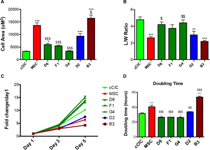
Figure 3.

Morphometric and proliferative characteristics of human CardioChimeras (hCCs). A, Cell surface area and (B) length to width (L/W) ratio of the parent cells and hCCs. C, Proliferation rate of parent cells and hCCs represented as fold change over day 1, and (D) cell doubling time represented in hours. N=3 independent experiments. Statistical analysis was performed by 1‐way ANOVA multiple comparison with Dunnett. *P<0.05 vs c‐kit+ cardiac interstitial cell (cCIC), **P<0.001 vs cCIC, ***P<0.0001 vs cCIC, $ P<0.05 vs mesenchymal stem cell (MSC), $$ P<0.001 vs MSC, $$$ P<0.0001 vs MSC. Error bars are ±SEM.
