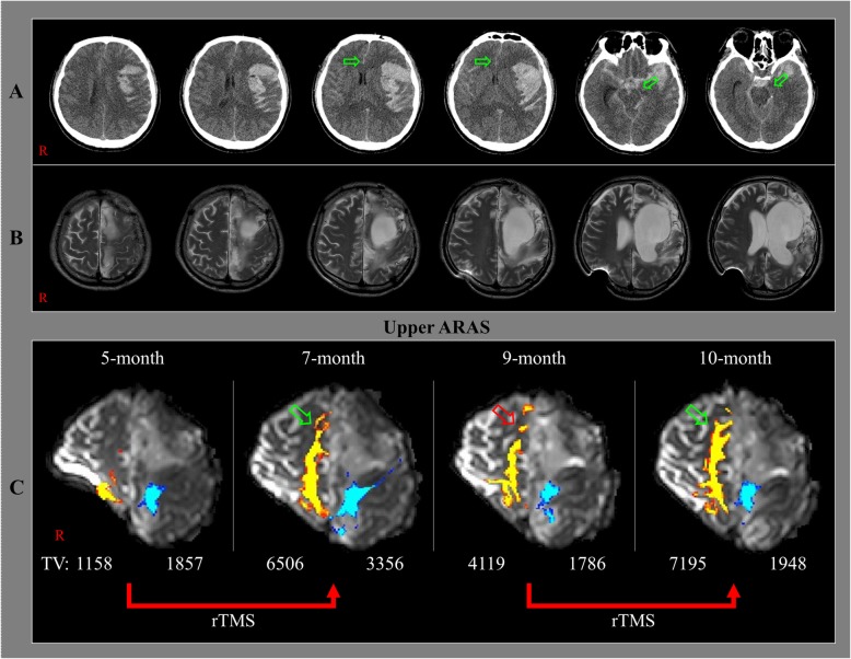
Fig. 1.
a Brain computed tomography images at onset show subfalcine and transtentorial herniations (arrows). b Brain magnetic resonance images at 5 months after onset show leukomalactic lesions in the left fronto-parieto-temporal lobes. c Results of diffusion tensor tractography (DTT) of the upper ascending reticular activating system (ARAS) of the patient. On the 7-month DTT, the neural tract of the right prefrontal lobe is increased (green arrow) compared to that on the 5-month DTT. However, the same neural tract is decreased (red arrow) on the 9-month DTT, but it is again increased (green arrow) on the 10-month DTT. ARAS: ascending reticular activating system, TV: tract volume, rTMS: repetitive transcranial magnetic stimulation

