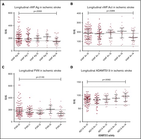
Figure 2.
Longitudinal patterns in ischemic stroke. Longitudinal changes in all haemostatic markers were measured at presentation (t0), 24 hours later (t1), 48 hours post presentation (t2), 5-7 days post presentation (t3) and final follow up from 6 weeks post presentation (t4). Median follow up time for ischaemic stroke specifically was 257 days (range 48-889). (A) Decrease in VWFAg from presentation (median 196.9 IU/dL) to final follow up (median 157.7 IU/dL) was observed (P = .0093 on matched paired testing). The same trend was seen with VWFAc from presentation (median 188.7 IU/dL) to final follow up (median 143.7IU/dL; P = .0289) (B), and FVIII (presentation median 178.6 to final follow up 137.7 IU/dL; P = .0149) (C). (D) A clear reverse trend was seen with ADAMTS13Ac in ischaemic stroke, demonstrating a significant increase in ADAMTS13Ac from presentation (median 85.9 IU/dL) to final follow up (median 96.8IU/dL, P = .0092).

