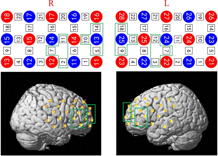
FIGURE 3.
Four regions of interest (ROIs): the frontal superior left area (Frontal_Sup_L, which contains channels 9, 13, and 18), the frontal inferior left area (Frontal_Inf_L, which contains channels 3, 7, and 12), the frontal superior right area (Frontal_Sup_R, which contains channels 5, 10, and 14), and the frontal inferior area right (Frontal_Inf_R, which contains channels 2, 7, and 11) were classified according to the existing anatomical calibration system, and related research results. L: left. R: right.

