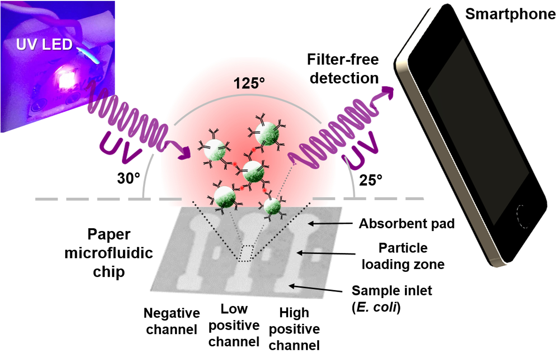
Figure 1.

Schematic illustration of the UV LED enhanced particle immunogglutination assay on paper microfluidics and subsequent smartphone detection. Antibodies (Y-shaped) are conjugated to the green fluorescent polystyrene particles (green spheres). The presence of E. coli antigens (red dots) triggers antibody-antigen binding and subsequently immunoagglutination of particles.
