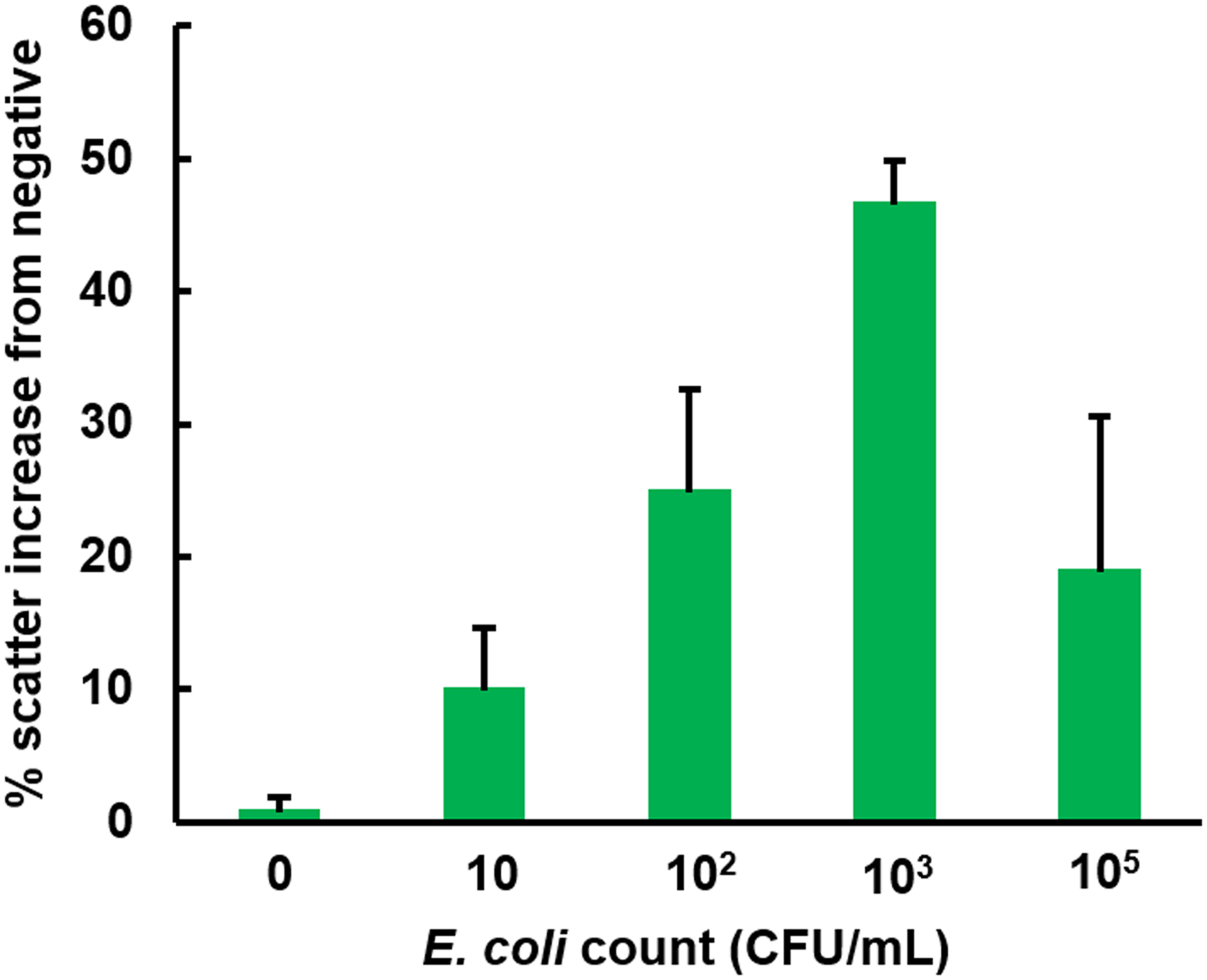
Figure 4.

The result of paper microfluidic assay for the E. coli in deionized water using the 385 nm UV LED. Green pixel intensities were evaluated and double-normalized as described in Materials and Method. 920 nm anti-E. coli conjugated PS particles were pre-loaded to the center of each paper microfluidic channel prior to the assays. Average of three different experiments, each time using different samples and different paper microfluidic chips. Error bars are standard errors.
