. 2019 Sep 27;10(42):9650–9662. doi: 10.1039/c9sc02886b

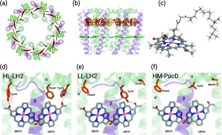
Fig. 1. (a) and (b) Structure of LH2 complex from Rps. acidophila. The LH2 complexes are circular structures formed by nine dimeric units, the α and β chains, binding one catotenoid molecule (not shown) and three bacteriochlorophyll a (BChl) molecules (α and βBChl, and B800). (c) Representation of the βBChl extracted from the LL–LH2 crystal structure. The atoms represented as spheres were included in the QM part in the multiscale calculations (see Methods), whereas the other atoms were described with the MMPol method. (d, e and f) Schematic representation of the local protein environment for α and βBChl within the same αβ unit inside the three complexes: HL–LH2, LL–LH2 and HM–PucD, respectively.

Fig. 1