
Figure 4.
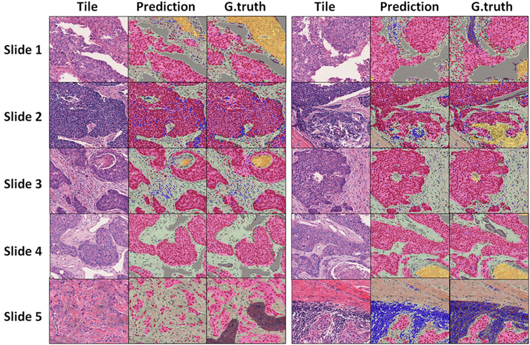
Qualitative examination of segmentation results on the testing set. Representative tiles from each the testing set. Slide 1 (right) and Slide 5 (right): Non-cellular components of “lymphocyte regions” (grey) were present in ground truth (for training) but were mapped to stroma in output. Slide 2 (left): enclosed stromal region within a tumor nest is missed in ground truth but is picked up by trained model. Slide 2 (right) and Slide 3 (right) algorithm misclassified small necrotic region as stroma. Slide 5 (left): Ground truth connects small, scattered tumor nests under one tumor “region”, whereas the model learns to more accurately delineate region boundaries.