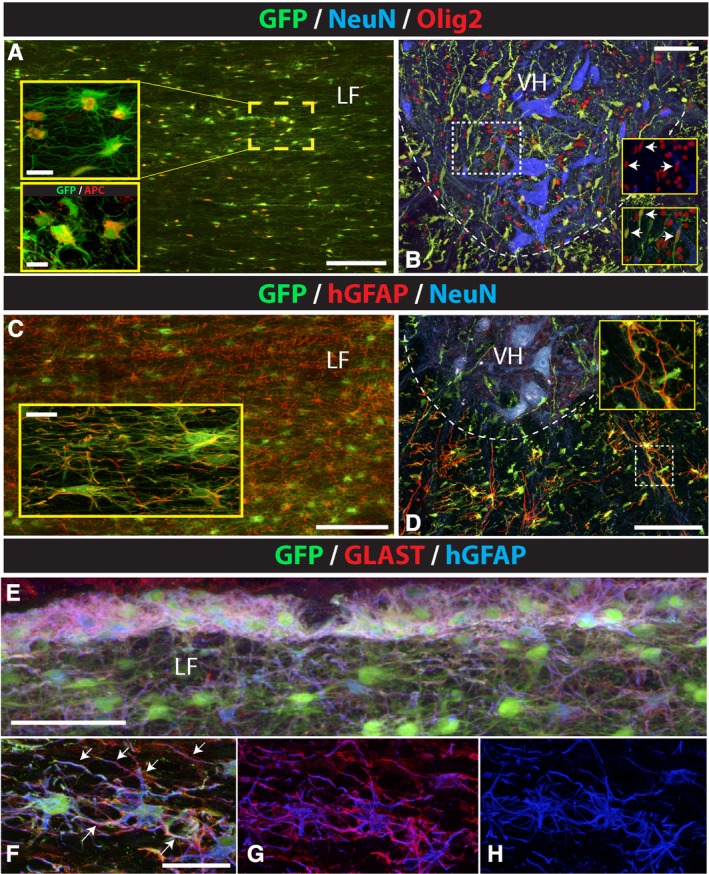
Figure 3.

Preferential astrocyte and oligodendrocyte differentiation of subpially injected hNSCs. A‐D, Analysis of horizontal and transverse spinal cord section stained with GFP/Olig2, GFP/hGFAP, or triple‐stained with GFP/NeuN/Olig2 and GFP/NeuN/hGFAP antibodies show a high density of Olig2‐immunoreactive cells co‐localizing with GFP in the white and gray matter in cervical segments (A, B). Staining with APC antibody shows expected cytoplasmic expression in GFP+ cells (A, lower inset). hGFAP expression was seen in astrocytes in the white matter but not in GFP+ cells in the gray matter (C, D). E‐H, Co‐staining of horizontal spinal cord sections with GFP/GLAST and hGFAP antibody showed that virtually all human astrocytes in the glia limitans as well as in the deeper white matter were GLAST positive. Scale bars: A, 150 μm, A (insets), 15 μm, 10 μm; B, 100 μm; C, 150 μm, C (inset), 15 μm; D, 100 μm; E, 60 μm; F, 30 μm; LF, lateral funiculus; VH, ventral horn
