Figure 3.
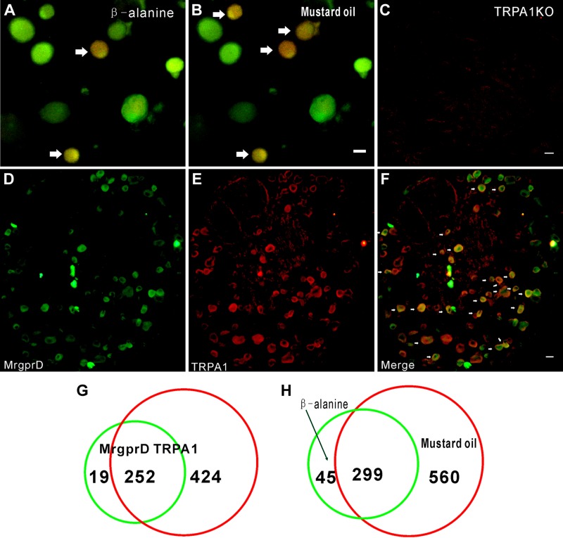
Most MrgprD+ DRG neurons are TRP-A1+. A, B) Representative images show DRG neurons responding to β-alanine (8 mM) and mustard oil (400 μM) in calcium imaging. Arrows: coresponsive cells. C) Immunostaining shows negative signal of TRP-A1 in DRG neurons from TRP-A1 KO mice. D–F) Immunofluorescence staining shows representative images of MrgprD+ cells (D), TRP-A1+ cells (E), and a merger of MrgprD+ and TRP-A1+ neurons (F). Arrows: double-labeled cells. G) Of 271 cells, 252 (92.9%) of MrgprD+ cells were TRP-A1+. H) Of 344 cells, 299 β-alanine (8 mM)–sensitive DRG neurons were coresponsive to mustard oil (400 μM). Scale bars, 20 µm.

