Figure 5.
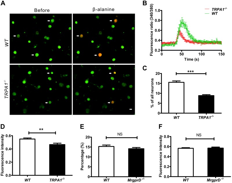
The β-alanine–induced response is decreased in TRP-A1-knockout mice. A) Fluorescent images of intracellular calcium flux induced by β-alanine. Arrows: neurons responsive to β-alanine in WT and TRPA1−/− DRGs. Scale bar, 20 µm. B) Representative fura-2 ratiometric responses in cultured DRG neurons. The curves show the responses of DRG neurons to β-alanine. C) The percentages of β-alanine–responsive neurons at the level of L4 DRGs from WT and TRPA1−/− mice (% of total neurons, n = 3 per group). D) The fluorescence intensities of β-alanine (8 mM)–induced calcium influx in DRG neurons of WT and TRPA1−/− mice (n = 3 per group). E) Percentages of mustard oil–responsive DRG neurons of WT and MrgprD−/− mice (% of total neurons, n = 3 per group). F) The fluorescence intensities of mustard oil–induced calcium influx in DRG neurons from WT and MrgprD−/− mice (n = 3 per group). ***P < 0.001, by independent samples t tests.

