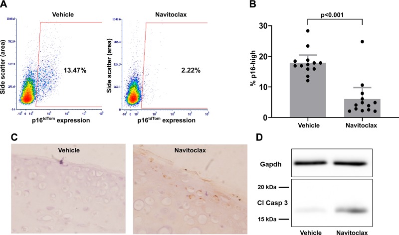
Figure 5.
Selective elimination of p16-high chondrocytes with navitoclax treatment. A) Representative flow plots showing the percentage of p16-high cells after a 3-d treatment with either DMSO vehicle control or 5 µM navitoclax after 3 wk of explant culture in senescence-inducing conditions. B) Quantification of p16-high cells in matched explants from 13 mice (1 hip DMSO control; 1 hip navitoclax/mouse). C) Immunohistochemistry for cleaved caspase-3 (Cl Casp 3; brown staining) and hematoxylin (blue staining) in paraffin-embedded explants fixed 18 h after treatment with DMSO vehicle control or navitoclax. D) Representative Western blot for Gapdh and cleaved caspase-3 using protein from hip cartilage explants harvested at 18 h after treatment with DMSO control or navitoclax.

