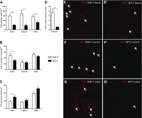
Figure 2.
KHK−/− and WT mice were fed 20% fructose (KHK-F and WT-F, respectively) for 8 wk. A–C) Distribution of CCK- (A), GLP-1- (B), and PYY-positive EECs (C) per section area (square millimeters) of mucosa in the ileum, cecum, and colon of KHK-F and WT-F. D) CCK peptide content in cecum tissue of KHK-F and WT-F. E–G′) Representative immunofluorescence staining of CCK-positive cells (red) in ileum (E, E′), cecum (F, F′), and colon (G, G′) of KHK-F (E, F, G) and WT-F (E′, F′, G′) mice (n = 5–6/group). Original magnification value, ×158. All values are means ± sem. Means were compared with Student’s t test. *P < 0.05, **P < 0.01, ***P < 0.001.

