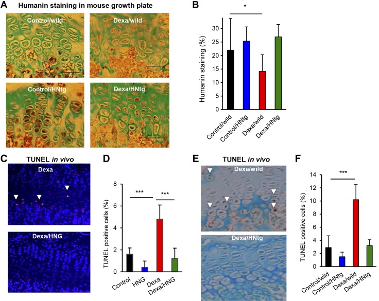
Figure 3.
Local (ex vivo) and systemic antiapoptotic effects of HN. A, B) Quantitative immunohistochemistry of HN expression (brown staining) with Alcian blue counter staining in the growth plate of wild-type and HNtg mice (C57BL6 background) treated with/without Dexa (2.5 mg/kg body weight/d) for 28 consecutive days. n = 3–6; scale bars, 50 µm; original magnification, ×40. C, D) Quantitative TUNEL analysis in growth plate of FVB mice treated with HNG (n = 5), fluorescent staining of apoptotic cells is shown in red and DAPI-staining of nuclei in blue. E, F) Quantitative TUNEL analysis in growth plate of HNtg animals (n = 3–6) treated with Dexa (2.5 mg/kg body weight), HNG (100 µg/kg body weight), or both, for 28 consecutive days. Apoptotic cells stained brown (DAB) and marked with white arrows. All error bars indicate sd. For statistical analysis, 1-way ANOVA was performed. Original magnification (C, E), ×20. *P < 0.05, ***P < 0.001.

