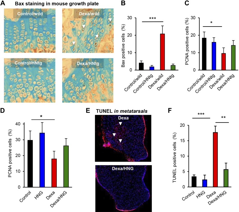
Figure 4.
Local and systemic effects on Bax, proliferation and apoptosis. A, B) Four-week-old male wild-type and HNtg mice (C57BL6 background, n = 3–6) were treated with Dexa (2.5 mg/kg body weight) for 28 consecutive days. Quantitative analysis for active Bax (brown staining, white arrows, original magnification, ×20) expression by using immunohistochemistry. C) PCNA analysis for cell proliferation in the growth plate of HNtg mice. D) PCNA analysis for cell proliferation in the growth plate of FVB mice (n = 5) treated with Dexa (2.5 mg/kg body weight/d), HNG (100 µg/kg body weight), or both, for 28 consecutive days. E, F) Fluorescent quantitative TUNEL in growth plates of fetal rat metatarsal bones (n = 5) treated with Dexa (1 µM), HNG (100 nM), or both, for 7 d. Apoptotic cells stained in red (white arrows) and nuclei in blue (DAPI). Original magnification (E), ×10. All error bars indicate sd. For statistical analysis, 1-way ANOVA was performed. *P < 0.05, **P < 0.01, ***P < 0.001.

