Figure 6.
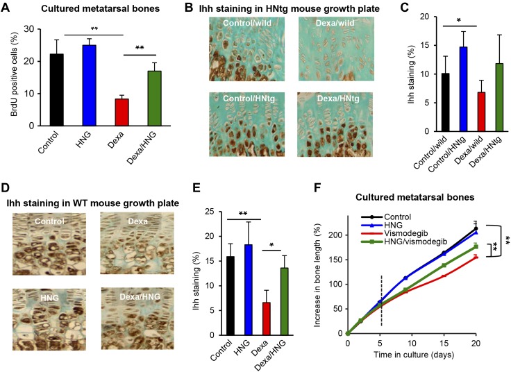
Effects of HN on Hh signaling. A) BrdU proliferation analysis in fetal rat metatarsal bones cultured ex vivo and treated with Dexa (10 µM), HNG (100 nM), or both, for 7 d (n = 5). B) Four-week-old male wild-type and HNtg mice (C56BL6 background, n = 3–6) were treated with Dexa (2.5 mg/kg body weight/d) for 28 consecutive days. Immunohistochemistry was performed to detect any changes in Ihh expression (brown staining). C) Quantitative analysis of Ihh, staining expressed as percent of growth plate area. D) Four-week-old FVB wild-type mice (n = 5) treated with Dexa (2.5 mg/kg body weight/d), HNG (100 µg/kg body weight), or both, for 28 consecutive days. Immunohistochemistry was performed to detect any changes in Ihh expression (brown staining) after pharmacological treatment with HNG. E) Quantitative analysis of Ihh, staining expressed as percent of growth plate area (n = 5). F) Fetal rat metatarsal bones cultured ex vivo were treated with the Hh inhibitor vismodegib (100 nM), HNG (38 nM), or both, for 5 d and thereafter followed for 15 d as indicated by the dotted vertical line without vismodegib or HNG (n = 9). All error bars indicate sd. One-way ANOVA was performed. Original magnification (B, D), ×20. *P < 0.05, **P < 0.01.

