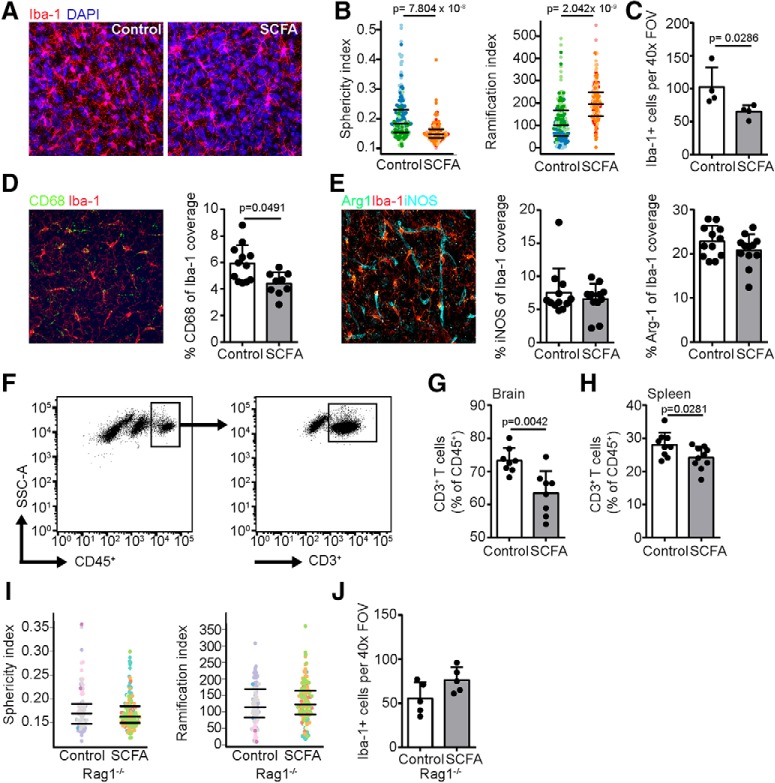
Figure 4.
Modulation of poststroke neuroinflammation by SCFA depends on peripheral lymphocytes. A, Representative maximum intensity projections of microglial staining using Iba-1 (red) and DAPI (blue) in the ipsilesional hemisphere 14 d after PT stroke with either control (left) or SCFA (right) supplementation. B, Microglial morphology was analyzed in 3D using an automated analysis algorithm in the ipsilesional cortex of mice 14 d after stroke, which revealed significantly reduced sphericity (left) and increased number of branch nodes (right) as markers of reduced microglial activation by SCFA compared with control treatment. Each symbol represents one microglia. Different colors group together microglia from the same mouse. C, Number of microglia found per 1 high-power (40×) FOV in the perilesional cortex. D, Coexpression coverage analysis in the ipsilateral hemispheric cortex for CD68 and Iba-1 expressed as percentage of Iba-1 from a maximum intensity projection. Representative immunofluorescence image (red represents microglia; green represents CD68) (left) and quantification (right). E, Coexpression coverage analysis in the ipsilateral hemispheric cortex for iNOS and Arginase1 (Arg1) with Iba-1 expressed as percentage of Iba-1 coverage area from a maximum intensity projection. Representative immunofluorescence image (red represents microglia; green represents Arg1; cyan represents iNOS) (left) and quantification (right). N = 3 mice per group and 3 images per hemisphere. In contrast to the effects of SCFA on microglia function, endothelial cells were unaffected by the SCFA treatment (Figure 4-1). F, Representative gating strategy for flow cytometric analysis of T cells (CD45+CD3+). SCFA supplementation significantly decreased the frequency of T cells in (G) brains and (H) spleens 14 d after stroke. N = 9 per group. Quantification of (I) sphericity (left) and ramification index (right) and (J) absolute cell counts of microglia 14 d after stroke in the perilesional cortex of Rag1−/− mice. In contrast to WT mice (compare with B,C), SCFA treatment did not affect microglia activation in lymphocyte-deficient Rag1−/− mice. All statistical analyses in this figure were performed using the Mann–Whitney U test.

