Figure 5.
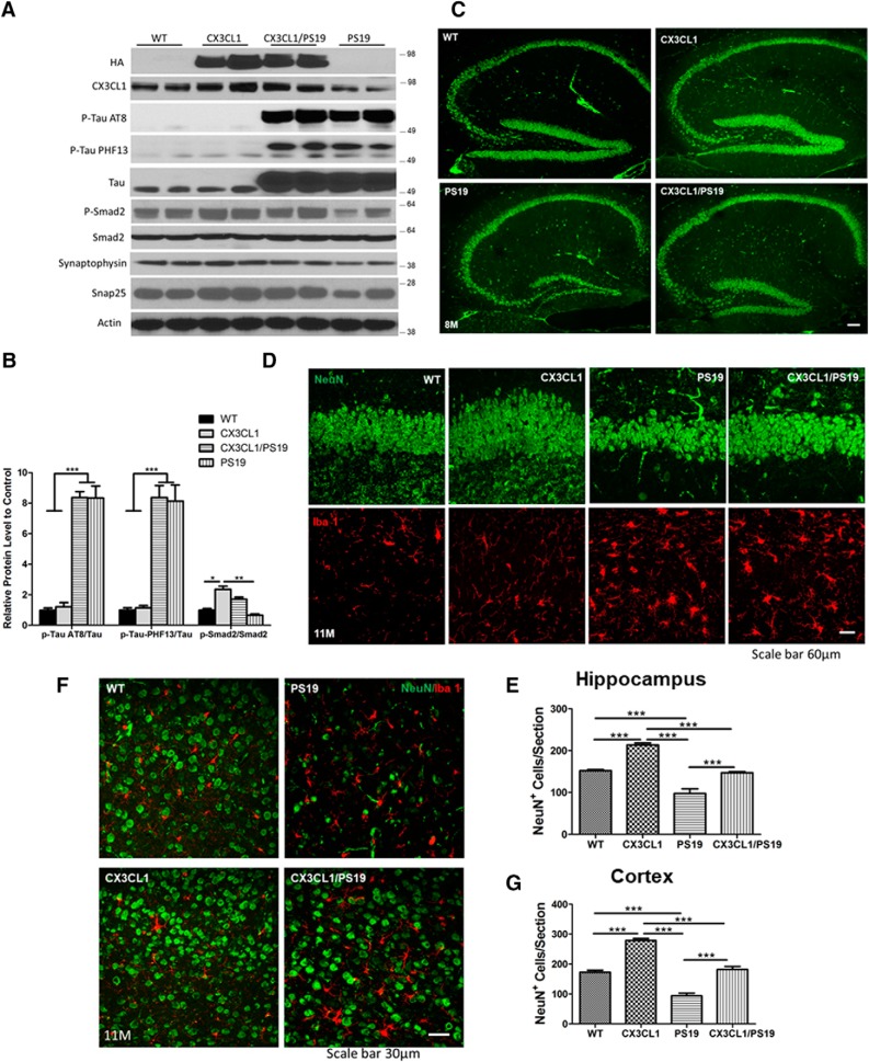
Overexpressed neuronal CX3CL1 rescues neuronal loss in PS19 mice. A, PS19 mice expressing mutant tau (P301S) in neurons. Western blot analyses from hippocampal lysates showed significantly elevated expression of total tau, and this elevation was maintained in Tg-CX3CL1/PS19 mice. Phosphorylated tau, detected by AT8 or PF13 antibodies, showed similar hyperphosphorylation in PS19 and Tg-CX3CL1/PS19 brains. Moreover, AT8+ punctae remained in Tg-CX3CL1/PS19 brains (Figure 5-1). CX3CL1 levels were lowered in PS19 brains, but were elevated due to CX3CL1 overexpression validated by two antibodies. p-Smad2 and synaptophysin levels were lower in PS19 mice, and this reduction was reversed by overexpression of CX3CL1. B, Bar graphs showed relative levels of AT8+ tau/tau, PHF13+ tau/tau, and p-Smad2/Smad2 (N = 2 in each group; three experiments; *p < 0.05, **p < 0.01, ***p < 0.001, two-way ANOVA with post hoc Bonferroni's test). C, Loss of neurons in CA and DG was visible in PS19 mice, and this reduction was partially rescued. D, Enlarged view of DG showing dramatic loss of neurons labeled by NeuN (green) in PS19 mice. Activation of microglia, labeled by Iba1 antibody, was evident in PS19 mice and also in Tg-CX3CL1/PS19 mice. F, Loss of neurons was visible in the cortical region of PS19 mice, and this reduction was significantly reversed after overexpression of CX3CL1 in Tg-CX3CL1/PS19 mice. Scale bar, 60 μm. E, G, Quantification of neurons in images of hippocampal (E) and cortical (G) regions, as specified (N = 3 slices each; *p < 0.05, **p < 0.01, ***p < 0.001, two-way ANOVA with post hoc Bonferroni's test).

