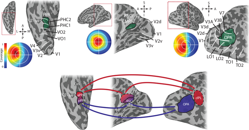
Figure 1. Scene-selective cortical regions.
A) Group average data showing the location of the scene-selective cortical regions with respect to anatomy and retinotopically-defined areas. The circular insets show the portion of the visual field eliciting the strongest response within each scene region based on population receptive field mapping. PPA (left) is located in and around the collateral sulcus on the medial part of the ventral temporal cortex. It overlaps with retinotopically defined regions PHC-1, PHC-2 and VO-2 and responds most strongly to stimuli in the contralateral upper visual field. RSC/MPA (middle) is located in medial parietal cortex in and around the ventral portion of the parieto-occipital sulcus. It responds most strongly to stimuli in the contralateral visual field with no clear bias to the upper or lower visual field. OPA (right) is located near the transverse occipital sulcus in occipito-parietal cortex. It overlaps most prominently with V3B and LO2, but also with V3A, V7/IPS0, and LO1, and responds most strongly to stimuli in the contralateral lower visual field.
B) Relationship between the different regions based on functional connectivity. There is strong functional connectivity between all three regions, but posterior PPA (pPPA) shows stronger connectivity to OPA and posterior parts of RSC/MPA, while anterior PPA (aPPA) shows stronger connectivity with the caudal inferior parietal lobe (cIPL), a region anterior to OPA, and anterior parts of RSC/MPA as well as adjoining regions in posterior cingulate cortex. This pattern of functional connectivity might reflect separate networks for perceptual (pPPA, OPA, posterior RSC/MPA) and memory-based (aPPA, cIPL, anterior RSC/MPA) processing. COS – collateral sulcus, OTS – occipitotemporal sulcus, MFS – mid-fusiform sulcus, CaS – calcarine sulcus, POS – parieto-occipital sulcus, IPS – intraparietal sulcus

