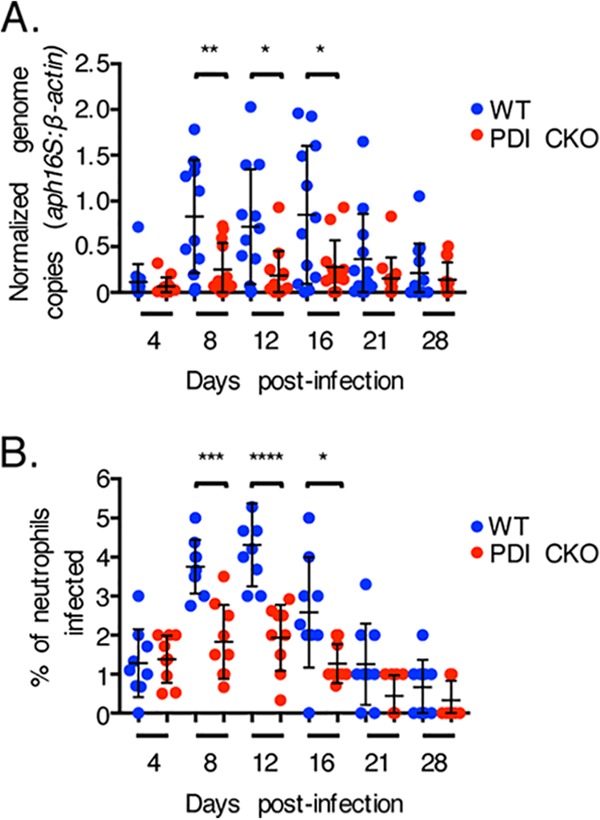
FIG 2.

PDI is critical for A. phagocytophilum to productively infect mice. PDI CKO or wild-type (WT) mice were infected with A. phagocytophilum DC organisms. (A) Peripheral blood drawn on the indicated days postinfection was analyzed by qPCR using gene-specific primers. Relative A. phagocytophilum 16S rRNA gene (aph16S)-to-murine β-actin DNA levels were determined using the cycle threshold (2−ΔΔCT) method. Data are the mean normalized bacterial loads ± SD calculated for 12 WT and 12 PDI CKO mice. (B) Peripheral blood samples were also examined for ApV-containing neutrophils by light microscopy. Each dot corresponds to the percentage of A. phagocytophilum-infected neutrophils as determined by examining at least 100 neutrophils per mouse. Data are the mean percentages ± SD determined for nine mice per group. Error bars indicate standard deviations among the samples per time point. Statistically significant values are indicated. *, P < 0.05; **, P < 0.01; ***, P < 0.001; ****, P < 0.0001.
