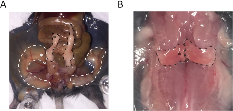
Figure 1: Anatomy of subcutaneous white adipose tissue (sWAT), epididymal white adipose tissue (eWAT), and brown adipose tissue (BAT) indicates regions used for collecting the samples.
(A) The regions outlined by white dashed lines represent sWAT, and the white asterisk highlighted location is the site for collecting sWAT. Regions outlined by black dashed lines are eWAT, and the black asterisk highlighted location is the site for collecting eWAT. (B) The regions outlined by black dashed lines are BAT, and the tissue collection region is highlighted by the asterisk.

