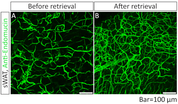
Figure 3: Comparison of IF images of blood vessels acquiredwith or without optical clearing step with 90% glycerol.
(A) Whole-mount immunofluorescence (IF) staining with anti-endomucin antibody (green) in sWAT. The sample was not subjected to the optical clearing step (step 3.10). (B) Whole-mount IF staining with anti-endomucin antibody in sWAT. The sample was subjected to the optical clearing step (step 3.10) before the mounting steps (steps 3.11 and 3.12).

