
Figure 1.
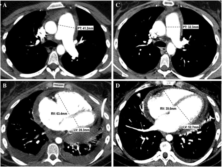
Chest computed tomography scans of our patient at diagnosis (A, B) and 6 months after treatment (C, D). Top row (A, C): Transverse computed tomography sections obtained at the level of the pulmonary trunk (A) and cardiac cavities (C) showing dilatation of the pulmonary trunk (41.2 mm) and right ventricular enlargement (63.4 mm) with a right ventricle/left ventricle ratio >1. Note the additional presence of pericardial and pleural effusion. Bottom row (B, D): Same anatomical levels as those shown on the top row, obtained 6 months later. Note the dramatic reduction in size of the pulmonary trunk (32.3 mm) (B) and right ventricle (39.6 mm) with normalization of the RV/LV ratio (D). Improvement of pericardial and left pleural effusion; resolution of right pleural effusion. PT, pulmonary trunk; RV, right ventricle; LV, left ventricle.