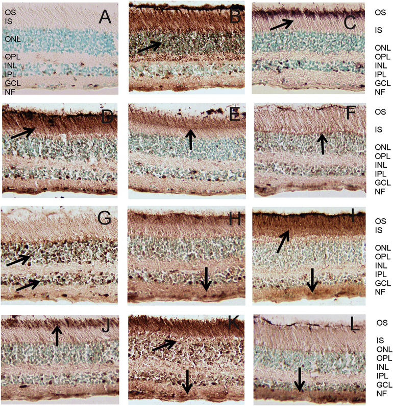
Figure 1.
Patterns of immunostaining of human retina cryosections with serum anti-retinal autoantibodies. Immunoperoxidase staining depended on antibody specificities and accessibility to the antigen in the tissue : (A) control staining, no serum, only secondary antibodies; (B) labeling of Ph layer; (C) labeling of OS in photoreceptor cells; (D) labeling of OS and IS in photoreceptor cells and GCL and NF; (E) labeling of OS and the junction between the IS and OS of rod and cone photoreceptors (F) labeling of OLM; (G) labeling of ONL and INL; (H) labeling of Ph cells, INL, IPL, GCL; (I) labeling of OS and IS and IPL and GCL; (J) labeling of OS of rods and cones, and outer limiting membrane; (K) Labeling of Ph cells, INL and GCL; and (L) labeling of IPL and GCL; Retinal layers are: OS, outer segments; IS, inner segments; Ph, photoreceptors; OLM, outer limiting membrane; ONL, outer nuclear layer; OPL, outer plexiform layer; INL, inner nuclear layer; IPL, inner plexiform layer; and GCL, ganglion cell layer; NF, nerve fiber layer. Arrows point at strongest immunostaining.

